







Short answer
A bone graft rebuilds jawbone that has thinned so a dental implant has solid bone to hold it. Graft material, from your own bone, a human donor, an animal source or a lab, is placed at the thin site and left to fuse with your jaw, usually over several months, before the implant goes in. A 3D scan shows whether you need one.
Why does the jawbone shrink after a tooth is lost?
The bone that held a tooth thins once the tooth is gone. A 2021 systematic review and meta-analysis found that when a tooth socket healed without a graft, the ridge at non-molar sites lost on average 2.73 mm of width and 1.71 mm of height at the outer wall, measured clinically.
On X-ray, the same review measured an average width loss of 2.54 mm at non-molar sites and 3.61 mm at molar sites, and concluded that the amount lost depends on the type of tooth. These are averages after a socket heals by itself, so an individual jaw can lose more or less.
The upper back jaw has a second problem. The American Association of Oral and Maxillofacial Surgeons explains that losing teeth deep in the upper jaw thins the bone, and the maxillary sinus, an air-filled space above the upper teeth, can expand downward into that space. That can leave too little bone height for an implant, so the location of the thin bone matters as well as the amount lost.
When do you need a bone graft for a dental implant?
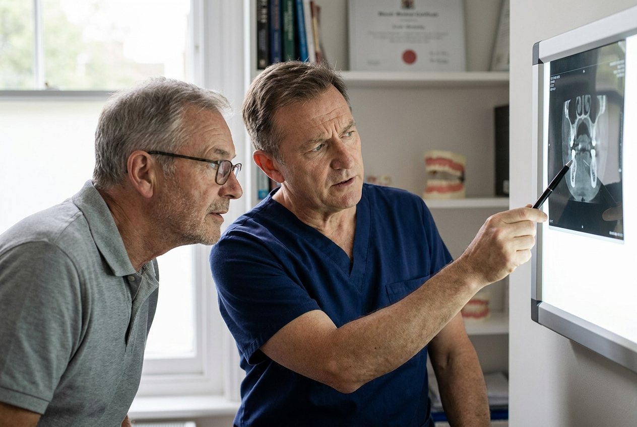
You need a bone graft when a 3D scan shows too little bone height or width to hold an implant firmly, or when a tooth is being removed and you want to keep the ridge for a future implant. If the scan shows enough bone, no graft is needed. The goal of grafting is good quantity and quality of bone at the site.
ANEWU describes three kinds of graft before an implant. Socket preservation places graft material into the socket when a tooth is removed, so the ridge keeps its shape. Ridge augmentation adds material along the outside of the jaw to rebuild width, height, or both. A sinus lift raises the sinus floor in the upper back jaw. Our page on bone grafting for dental implants covers each one.
Socket preservation has trial evidence behind it. A 2019 systematic review and meta-analysis of 22 randomized trials found that grafting the socket prevented 1.99 mm of horizontal bone loss compared with extraction alone, when sites were compared after a 3 to 6 month healing period. It could not say which grafting approach worked better. A graft will not suit every jaw: not every jaw can be rebuilt to the volume a standard implant needs.
Can you still get a dental implant without a bone graft?
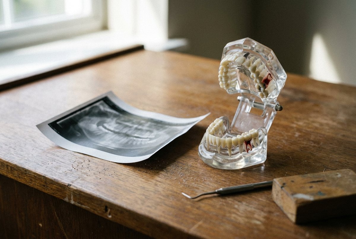
Sometimes. If your scan shows enough bone, a standard implant needs no graft. When bone is limited, short implants, tilted implants, zygomatic implants and pterygoid implants can avoid or reduce grafting for some people. None of them suits everyone, and each depends on your anatomy, which a 3D scan shows.
Short and tilted implants have been compared with the usual alternatives. A 2024 systematic review of 19 randomized trials (2,214 implants) found moderate to high certainty evidence of similar 5-year survival for 6-mm implants in the upper jaw instead of a sinus lift, but inconclusive or insufficient evidence for 4-mm and 5-mm implants used that way. A 2021 meta-analysis of 11 studies found no statistically significant difference in survival between tilted and straight implants in thin, fully toothless upper jaws after three years. Its authors describe tilting as a way to use the bone that is there without a sinus lift. The studies attached teeth soon after surgery (immediate loading); teeth attached that soon are provisional, and the final set comes after healing.
Zygomatic implants anchor in the cheekbone, and pterygoid implants anchor in dense bone at the back of the upper jaw. ANEWU describes both as options for specific anatomy and says not every upper jaw suits them. A 2023 systematic review of 18 studies of zygomatic implants (1,349 implants in 623 patients) found 96.2% survival at 6 years, in studies with at least 3 years of follow-up. Sinusitis was the most frequent biological complication, with a prevalence of 14.2% at 5 years.
What are the types of bone graft material?
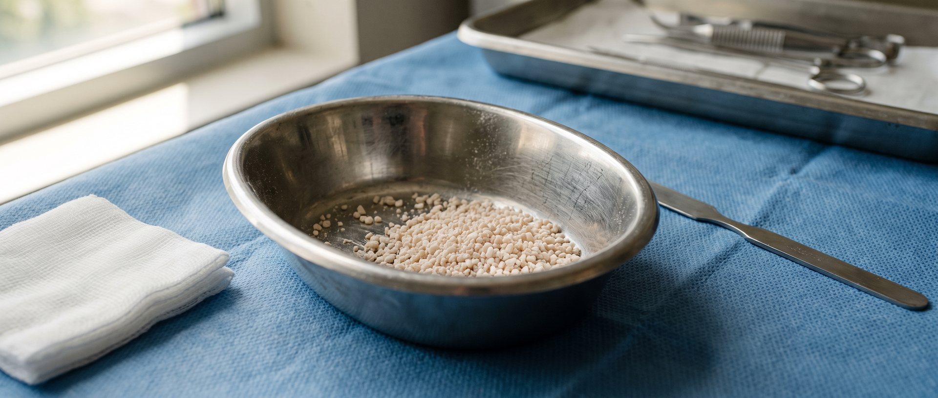
Four sources are used. Autograft is your own bone, allograft is screened human donor bone, xenograft is screened animal bone, and synthetic grafts are made in a lab. A graft can directly replace missing bone or act as a scaffold on which your own bone grows, and your oral and maxillofacial surgeon helps you choose the kind to use.
Each has trade-offs. The American Association of Oral and Maxillofacial Surgeons notes that your own bone carries no disease-transmission risk, but it means a second surgical site, most often the jaw, hip or lower leg. A 2026 review in the Journal of Periodontal Research adds that autografts supply living cells but can resorb unpredictably, while processed xenografts hold their volume well, and processed allografts and xenografts alike have less ability to prompt new bone.
Materials are often combined. The same review says blends of small amounts of the patient's own bone with slow-resorbing xenograft or synthetic material, protected by a membrane or mesh, improve contour stability in sinus lifts and in lateral and vertical bone augmentation. It also calls the ideal material choice controversial.
| Graft type | Where it comes from | Main advantage | Main drawback |
|---|---|---|---|
| Own bone (autograft) | Your jaw, hip or lower leg | No disease-transmission risk; living cells | A second surgical site; can resorb unpredictably |
| Human donor bone (allograft) | Screened and treated donor tissue | No second surgical site | Less able to prompt new bone |
| Animal-derived bone (xenograft) | Screened and treated animal tissue | Holds its volume; no second surgical site | Less able to prompt new bone |
| Synthetic (alloplast) | Lab-made, such as hydroxyapatite or tricalcium phosphate | No donor risk; lower cost | Growth may be less predictable |
How does a bone graft rebuild the ridge, step by step?
A bone graft rebuilds the ridge in five steps: a 3D scan maps the missing bone, the site is numbed and opened, graft material is placed, the graft fuses with your jaw over several months, and a follow-up scan confirms the site is ready before the implant is placed.
Healing time varies. In the 2019 socket-grafting meta-analysis, sites were compared after a 3 to 6 month healing period. For sinus lifts, a 2026 review describes a 2021 network meta-analysis of 34 randomized trials (842 two-stage lateral window lifts) that pooled trials with a "normal" healing period of 5 to 8 months. It compared graft materials, not healing times, so treat the range as typical of the research, not a rule. A bigger graft takes longer than a small socket graft, and ANEWU uses the second scan rather than a fixed calendar. For a sinus lift, AAOMS says the implant can sometimes be placed at the same time as the graft.
- Step 1, scan and plan: a 3D scan measures how much bone is present and where it is missing.
- Step 2, numb and open: the site is numbed, with sedation when appropriate, and the gum is opened over the thin area.
- Step 3, place the graft: material goes where bone is missing, sometimes under a membrane that keeps unwanted tissue out, and the gum is closed over it.
- Step 4, heal and fuse: over the next few months the graft integrates with your jawbone and hardens into a base an implant can hold.
- Step 5, confirm and place the implant: a follow-up 3D scan checks the site before implant placement is scheduled.
How is a sinus lift done for a dental implant?
A sinus lift adds bone between the upper jaw and the maxillary sinus so an implant has enough height. The surgeon lifts the sinus lining and places graft material beneath it, either through a small window cut in the side of the jawbone (the lateral window technique) or through a small hole made in the jawbone below the sinus (the vertical osteotome technique, also called transalveolar).
AAOMS describes both. In the lateral window technique an incision exposes the jawbone, a small window is cut into the bone, the sinus lining is lifted, graft material is placed, and the gum is sutured. In the vertical osteotome technique a small hole is made in the bone below the sinus, and an instrument called an osteotome lifts the lining so graft material can fill the space. AAOMS says a sinus lift may be done under local anesthesia, with sedation if necessary.
Survival is high but not uniform. A 2018 systematic review and meta-analysis of 17 clinical trials (637 patients, 1,610 implants) found 97.7% implant survival at 3 to 6 years after a sinus lift by either approach. Only one of the 17 was randomized; the other 16 were prospective non-randomized studies, and results varied a lot between them. Smoking was linked to worse survival in two studies (relative risk 4.8, with a wide confidence interval of 1.2 to 19.4), and the certainty of that evidence was rated very low to moderate. A 2019 European consensus report concluded that the lateral window sinus lift is reliable in the long term for the partly and fully toothless upper jaw.
Video transcript
A silent animation in five steps showing a sinus lift. Step 1: there is not enough bone height between the upper jaw and the sinus for an implant. Step 2: the surgeon lifts the sinus lining. Step 3: graft material goes beneath the lining. Step 4: the graft heals and joins your jawbone over time. Step 5: an implant is placed when the site is ready. The illustration is general. Your surgeon decides the technique and the timing from your scan.
How painful is a bone graft, and what are the downsides?
Expect soreness and swelling that peak early and then ease, plus some risk of complications. In a 2018 systematic review of 11 studies of lateral window sinus lift surgery, patient-reported discomfort peaked on day 1 and generally declined after that. A 2026 meta-analysis of maxillary bone regeneration pooled complication rates from 7.98% for sinus lifts to 26.44% for guided bone regeneration, with wide confidence intervals.
The pain data cited here come from sinus lifts, so that is what can be quoted. The 2018 review gathered patient-reported outcomes after the lateral window approach, and it found that severe discomfort occurred but not in most patients. The studies used different pain scales and time points, so there is no single pain score. ANEWU lists swelling and soreness for the first several days, soft foods and a follow-up visit, and says soreness settles well before the bone is ready. Follow the written instructions from your surgeon; our post-op instructions page has the general guidelines.
The 2026 meta-analysis pooled 100 articles. The most frequent complications were membrane or graft exposure, wound dehiscence (the stitched gum reopening) and infection, and guided bone regeneration and onlay grafting carried a higher risk of membrane exposure and wound dehiscence. A 2019 European consensus report found lateral and vertical ridge augmentation effective, but noted that the rate of associated complications was high for both. Taking your own bone adds a second surgical site.
What does a bone graft look like at ANEWU?
At ANEWU, Dr. Abtin Shahriari, an oral surgeon specializing in implant dentistry, performs every bone graft and implant. Each graft is planned from a 3D scan taken at your first visit, the graft is placed in a single appointment with the site numbed and sedation when appropriate, and a second scan confirms the site is ready before the implant is scheduled.
The first consultation is free at all three Georgia offices, Buckhead, Cumming and Dawsonville, and the scan is reviewed with you on screen. ANEWU's page lists three graft materials, your own bone, donor bone or a synthetic material, and also performs zygomatic and pterygoid implants for suitable anatomy. Cost depends on how much bone is missing, so you get a written figure for your own case after the scan, and CareCredit and Proceed Finance are accepted.
The scan, not a general rule, decides your plan, so bring questions like these. If you would like to see your own anatomy and hear which route fits, you can book a free consultation.
- Which graft material do you recommend for my site, and why?
- Is the graft done first, or at the same time as the implant?
- How long should I wait before the implant, and how will we know the site is ready?
- Would a short, tilted, zygomatic or pterygoid implant suit my anatomy instead?
- What are the risks in my case, and how do smoking and vaping change them?
- What written aftercare will I get, and who do I call?
Sources and further reading
- Post-extraction dimensional changes: A systematic review and meta-analysis Journal of Clinical Periodontology, 2021
- Bone Grafting and Membranes American Association of Oral and Maxillofacial Surgeons, patient information page, accessed October 2026
- Sinus Augmentation: Preparing Sinuses for Dental Implants American Association of Oral and Maxillofacial Surgeons, patient information page, accessed October 2026
- Bone Grafts: Everything You Need to Know Journal of Periodontal Research, 2026
- Effect of alveolar ridge preservation interventions following tooth extraction: A systematic review and meta-analysis Journal of Clinical Periodontology, 2019
- Short (6 mm or less) compared with 10-mm or longer dental implants in different clinical scenarios: A systematic review of randomized clinical trials with meta-analysis, trial sequential analysis and quality of evidence grading Journal of Clinical Periodontology, 2024
- Clinical success between tilted and axial implants in edentulous maxilla: A systematic review and meta-analysis Journal of Indian Prosthodontic Society, 2021
- Long-term treatment outcomes with zygomatic implants: a systematic review and meta-analysis International Journal of Implant Dentistry, 2023
- Clinical Performance of Dental Implants Following Sinus Floor Augmentation: A Systematic Review and Meta-Analysis of Clinical Trials with at Least 3 Years of Follow-up International Journal of Oral & Maxillofacial Implants, 2018
- Regeneration of alveolar ridge defects. Consensus report of group 4 of the 15th European Workshop on Periodontology on Bone Regeneration Journal of Clinical Periodontology, 2019
- Patient-Reported Outcomes After Lateral Wall Sinus Floor Elevation: A Systematic Review Implant Dentistry, 2018
- Oral bone regeneration and associated complications: A systematic review and meta-analysis Journal of Stomatology, Oral and Maxillofacial Surgery, 2026
This article is general information about dental and facial procedures, not a diagnosis or medical advice. Every jaw and every health history is different, so the right plan for you can only be set after an exam and imaging with a surgeon.